
the elegant dinoflagellate: it’s toxic strength is in it’s numbers

Scanning electron micrographs reveals cell wall structure that is used to identify the species.
When my husband, Alfred R. Loeblich III, was an associate professor at Harvard, he was assisted by his first wife, Laurel Loeblich, PhD, and his students in studying dinoflagellates – these exquisite, sometimes glowing, often toxic, microorganisms. When found in congregations of large numbers in harmful algal blooms known as red tides, they can contaminate fish and in turn make a person very sick.
Alfred produced these gorgeous scanning electron micrographs of dinoflagellates, which originals I stumbled across when we FINALLY got around to cleaning out our basement which had been clogged with maybe 300 to 400 boxes of musty old reprints, journals (some dating back to the 1920’s !), books, chemicals, and various lab equipment.
Alfred and his collaborators produced hundreds of these scanning electron micrographs of about 20 or 30 different species. Sometimes they were cultured in a test tube, sometimes they were collected from nature (you can see diatoms in the background of the Gonyaulax acatenella). Pictured above are just 4 of them, which were all marine organisms. But he studied other freshwater species as well.
He has been published in Science and Nature magazines, along with publications by Phykos, Wiley, McGraw Hill, and more (see below).
Pictures and publications
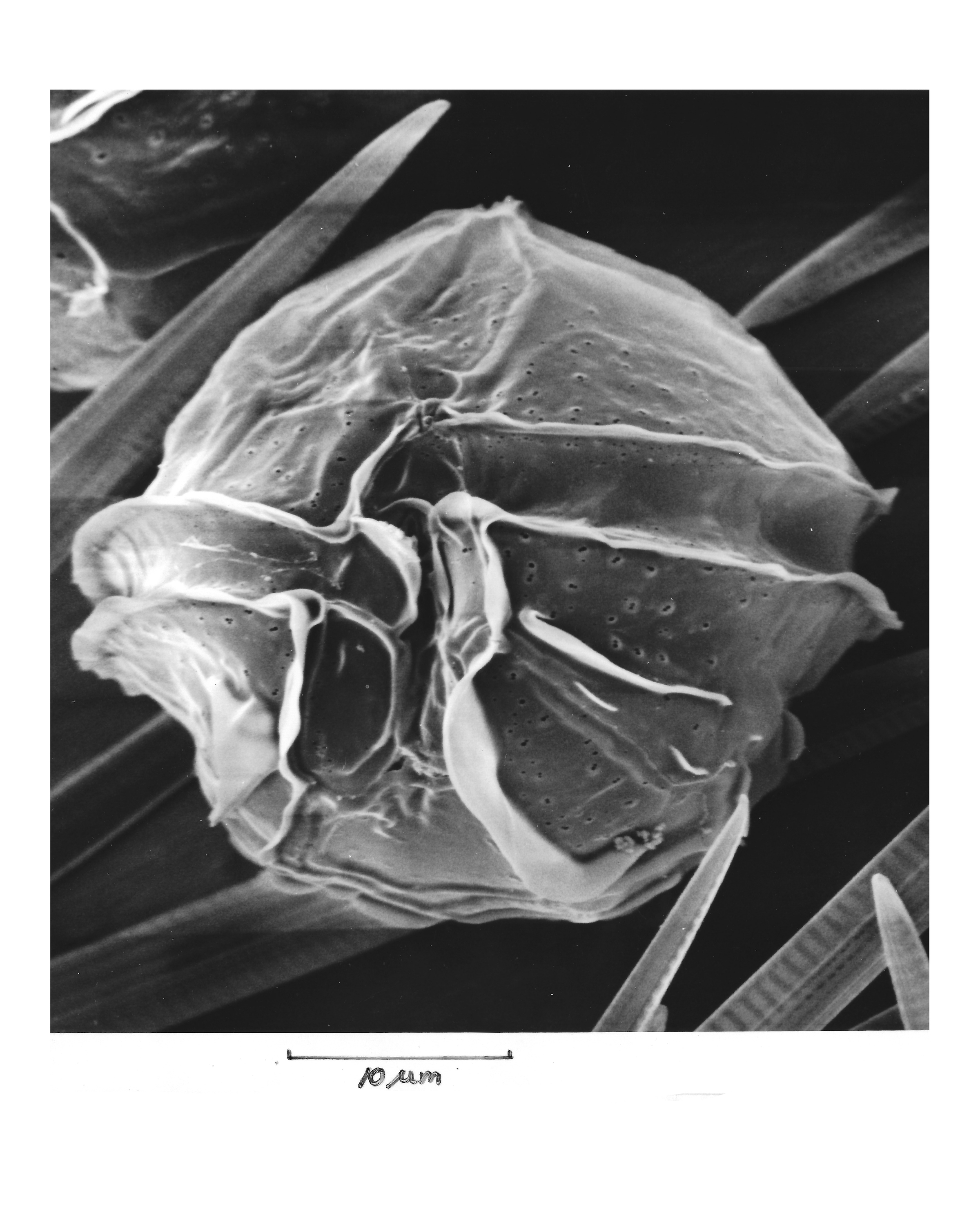
Ceratocorys horrida
published in Synopsis and Classification of Living Organisms, 1982, McGraw Hill Book Co. Inc., on plate 13. When the cell stops swimming it rests at bottom of test tube on its head (the anterior end of the cell). Isolate came from University of California at Santa Barbara.
Gonyaulax acatenella
collected from a red tide in Malaspina Inlet, B.C., Canada, June 10, 1965. Published in Proceedings of the First International Conference on Toxic Dinoflagellate Blooms, Nov. 1974. Published in 1975.
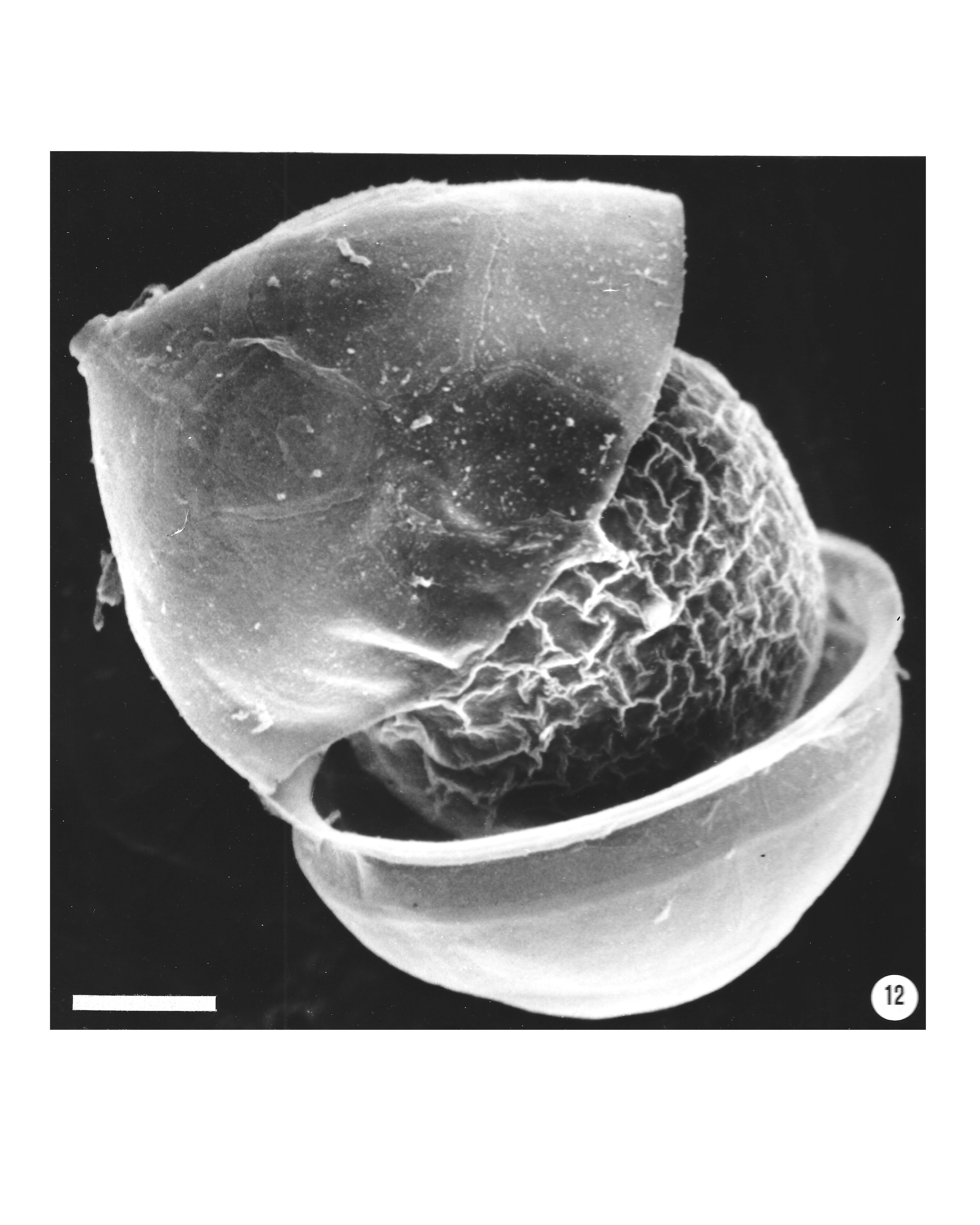
Peridinium trochoideum
published in Proceedings Biological Society of Washington, vol.89, p.284. Shows method of shedding the cell wall at the time of cell division. Culture was obtained from Woods Hole Oceanographic Institution, MA.
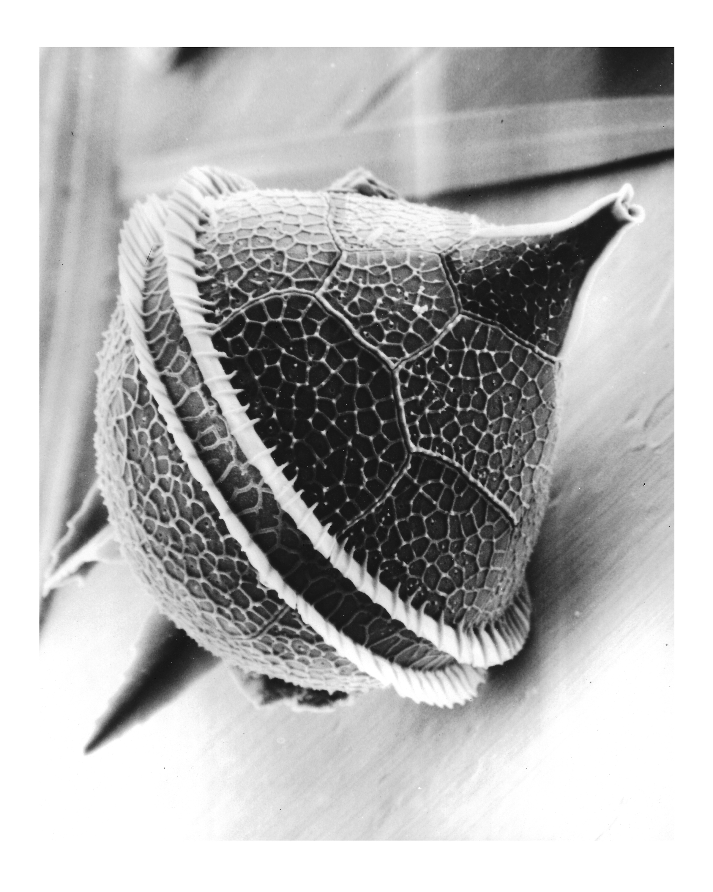
Protoperidinium steinii
published in Synopsis and Classification of Living Organisms, 1982, McGraw Hill Book Co. Inc., plate 14. Cell is from a plankton sample.
Learn more about dinoflagellates here:

Hi Wendy. Sorry I missed replying to you sooner! Quite impressive artic